The Journal of Clinical and Preventive Cardiology has moved to a new website. You are currently visiting the old
website of the journal. To access the latest content, please visit www.jcpconline.org.
Left Ventricular Cyst in a Young Female. What Could be the Cause?
Volume 4, Jul 2015
S. R. Mittal DM (Cardiology), Ajmer, India
J Clin Prev Cardiol 2015;4(3):76-77
A 20 years female was evaluated for atypical chest pain. Clinical examination, electrocardiogram and skiagram of chest were normal. Echocardiography revealed a cystic lesion in LV apex (Fig 1 – image 1 & 2). The cyst had intracavity echoes (arrow). Underlying myocardium was normal. There was mild mitral and tricuspid regurgitation (Fig. 1- image 3 & 4). There was no other lesion. Skiagram of chest and sonography of abdomen were normal. Total eosinophil count was 1050/cu mm. Casoni skin test was negative. Patient could not afford further evaluation. She was empirically advised albendazole. She did not come for follow up.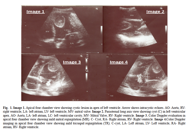
Discussion
Cystic lesions of heart are rare. Hydatid cyst is the commonest. Mostly it is associated with extracardiac lesions. Isolated cardiac involvement is extremely rare. Heterogeneous echogenicity with dense contents support possibility of hydatid cyst. Intracystic echogenic shadows could be due to scolices of echinococcus. Eosinphilia is a supportive findings. Positive Casoni test supports diagnosis but a negative test does not exclude the possibility.
Cardiac hemangioma can present as left ventricular cyst (1). Typically these are pedunculated, freely mobile and hyperechoic on echocardiography. Due to high vascularity, these tumors show rapid homogeneous enhancement during gadolinium infusion and a vascular blush on coronary angiography. Bronhcogenic cysts are typically intramyocardial with irregular border (2). There is no contrast enhancement. Histologically the cavity is characteristically lined by ciliated respiratory epithelium. Rarely left ventricular apical thrombus can have a cystic appearance (3). Underlying myocardium shows akinesia or dyskinesia. Electrocardiogram shows evidence of myocardial necrosis. Proper anticoagulation for one month usually reduces the size of a thrombus. Left ventricular myxoma can also undergo cystic degeneration. Myxomas are pedunculated. Freely mobile with irregular surface. Blood cysts are usually small and attached to endocardium (most often valvular endocardium). However, there are case reports of large left ventricular blood cyst (4).
Echocardiography has high sensitivity and specificity in identifying a cyst However, definite. etiological diagnosis may be difficult in presence of atypical location, calcification and heterogeneous echogenicity due to dense contents. Cardiac CT is useful in such situations. In case of doubt, cardiac MRI provides conclusive information (5).
References
- Fathala A. Left ventricular cyst; unusual echocardiographic appearance of a cardiac hemangioma. Circulation 2012;125:2171-2.
- Klass O, Haffmann MHK, Ludwig B, Leithauser F, Hannekum A. Left ventricular bronchogenic cyst. Circulation 2007;116:e385- e387.
- Ozlu MF, Ozean F, Tufekeioglu O. Left ventricular apical cystic thrombus mimicking a hydatid cyst. Can J Cardiol 2009;25:e266.
- Ohmoto Y Tsuchihashi K, Tanaka S, Shimamoto K, Iimura O. Giant endocardial blood cyst in left ventricle resected by transaortic valve approach. Chest 1993;103:965-6.
- Canpolal U, Yorgun H, Sunman H, Aytemir K. Cardiac hydatid cyst mimicking left ventricular aneurysm and diagnosed by magnetic resonance imaging. Turk Kardiyol Dem Ars- Arch Turk Soc Cardiol 2011;39:47-51
