The Journal of Clinical and Preventive Cardiology has moved to a new website. You are currently visiting the old
website of the journal. To access the latest content, please visit www.jcpconline.org.
Right Ventricular Mid-cavity Dynamic Systolic Obstruction due to Dyskinetic Interventricular Septum
Volume 4, Apr 2015
S. R. Mittal DM, Ajmer, India
J Clin Prev Cardiol 2015;4(2):50-1
A ten-year-old child was referred for echocardiography. He was diagnosed to have “heart trouble” at the age of three months following an episode of “bronchitis”.
He was asymptomatic on tablet digoxin 0.125 mg/ day, tablet frusemide 20 mg/day, tablet carvedilol 6.25 mg BD and tablet enalapril 15 mg BD. Clinical
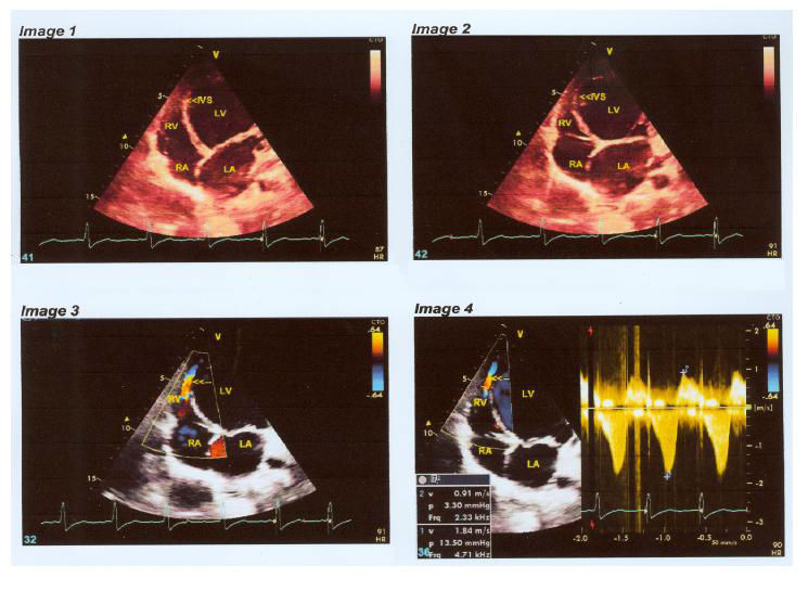
Figure 1. (Image 1): Apical four chamber view in diastole showing dilated LA and LV with thin IVS. IVS- interventircular septum, LA- left atrium, LV- left ventricle, RA- right atrium, RV- right ventricle.
(Image 2): Apical four chamber view in systole showing dyskinesia of interventricular septum (IVS).
(Image 3): Color Doppler image showing systolic turbulent flow between right ventricular (RV) apex and mid cavity adjacent to region of dyskinetic interventricular septum (IVS).
(Image 4): Doppler evaluation of the systolic turbulent flow showing velocity of 1.84 m/sec and gradient of 13.5 mm Hg.
examination revealed only mild leftward displacement of apical impulse. Electrocardiogram was within normal limits. Echocardiogram revealed dilated left ventricle. End diastolic dimension was 6.5cm. End systolic dimension was 5.3 cm. Interventricular septum was thin and infarcted. Other walls of left ventricle showed normal thickness and contractility. Left ventricular ejection fraction was 35%. Right ventricular functions were normal. Origin of left coronary artery was normal. Apical four chamber view revealed mild mitral regurgitation. Interventricular septum (IVS) was pushed toward right ventricle during systole (Figure 1- image 1 and 2). There was no aneurysm. Right ward systolic bulging of interventricular septum resulted in dynamic systolic obstruction in right ventricular mid cavity with turbulent flow (Figure 1 image 3). On Doppler evaluation peak systolic gradient across turbulence was 13.5 mm Hg (Figure 1 image 4). Gradient did not alter with any maneuver. Final echocardiographic diagnosis was viral myocarditis. It is hypothesized that rightward motion of dyskinetic IVS and normal inward movement of right ventricular free wall produced right ventricular mid cavity dynamic systolic gradient with turbulent flow. To the best of our knowledge, such an association has not been described (1,2).
References
He was asymptomatic on tablet digoxin 0.125 mg/ day, tablet frusemide 20 mg/day, tablet carvedilol 6.25 mg BD and tablet enalapril 15 mg BD. Clinical
Figure 1. (Image 1): Apical four chamber view in diastole showing dilated LA and LV with thin IVS. IVS- interventircular septum, LA- left atrium, LV- left ventricle, RA- right atrium, RV- right ventricle.
(Image 2): Apical four chamber view in systole showing dyskinesia of interventricular septum (IVS).
(Image 3): Color Doppler image showing systolic turbulent flow between right ventricular (RV) apex and mid cavity adjacent to region of dyskinetic interventricular septum (IVS).
(Image 4): Doppler evaluation of the systolic turbulent flow showing velocity of 1.84 m/sec and gradient of 13.5 mm Hg.
examination revealed only mild leftward displacement of apical impulse. Electrocardiogram was within normal limits. Echocardiogram revealed dilated left ventricle. End diastolic dimension was 6.5cm. End systolic dimension was 5.3 cm. Interventricular septum was thin and infarcted. Other walls of left ventricle showed normal thickness and contractility. Left ventricular ejection fraction was 35%. Right ventricular functions were normal. Origin of left coronary artery was normal. Apical four chamber view revealed mild mitral regurgitation. Interventricular septum (IVS) was pushed toward right ventricle during systole (Figure 1- image 1 and 2). There was no aneurysm. Right ward systolic bulging of interventricular septum resulted in dynamic systolic obstruction in right ventricular mid cavity with turbulent flow (Figure 1 image 3). On Doppler evaluation peak systolic gradient across turbulence was 13.5 mm Hg (Figure 1 image 4). Gradient did not alter with any maneuver. Final echocardiographic diagnosis was viral myocarditis. It is hypothesized that rightward motion of dyskinetic IVS and normal inward movement of right ventricular free wall produced right ventricular mid cavity dynamic systolic gradient with turbulent flow. To the best of our knowledge, such an association has not been described (1,2).
References
- Armstrong WF, Ryan T. Dilated Cardiomyopathies. In Feigenbaum’s Echocardiography. Armstrong WF, Ryan T (eds). Wolters Kluwer; Philadelphia. 2010:507-38.
- Gokhale R, Basra M, Vacanti V, et al. Echocardiographic assessment of non obstructive cardiomyopathies. In Comprehensive Textbook of Echocardiography Nanda NC (ed).Jaypee, New Delhi. 2014:1369-417.
