The Journal of Clinical and Preventive Cardiology has moved to a new website. You are currently visiting the old
website of the journal. To access the latest content, please visit www.jcpconline.org.
Hypertrophic Muscle Band in the Left Ventricle in an Asymptomatic Patient
Volume 3, Apr 2014
Ramit Wadhwa, MBBS, PGDCC, Hardeep Kaur Grewal, MBBS, PGDCC Rahul Mehrotra, MD, DNB, Ravi R Kasliwal, MD, DM Gurgaon, India.
J Clin Prev Caridiol. 2014;3(2):71-2
Introduction
Hypertrophic muscle band (HMB) in left ventricular (LV) cavity is a rare echocardiographic finding, especially in the absence of LV hypertrophy. Although this entity has been seen in the western population (1,2), it is rarely seen in their Asian counterparts. So far, to the best of our knowledge, only three cases have been reported from Asia. Two cases have been reported from Korea, one of a 5-year-old child (3) and another of a 25-year-old female (4), while one case has been reported from India - that of a 37-year-old male (5). This suggests that this entity is rare and can be seen in all age groups including adults and children. We hereby report a case of incidentally detected HMB on echocardiography.
Case
Our case was a 40-year-old lady who came to our hospital for routine health checkup. She was asymptomatic with no comorbidity and her physical examination was unremarkable. ECG findings were within normal limits.
Echocardiography revealed a prominent, hypertrophied muscle band in the LV, measuring approximately 1.6 cm in thickness, extending from basal anterior septum to apical LV free wall (Figs. 1 and 2). Echo texture was the same as that of inter ventricular septum. LV wall thickness was within normal limits from base to apex and LV contractility was also normal. No significant resting or provocable intracavity gradients were noted. Twenty-four hour Holter monitoring was also done, and it did not reveal any arrhythmia.
.jpg)
Figure 1. Parasternal long axis view with hypertrophied muscle band.
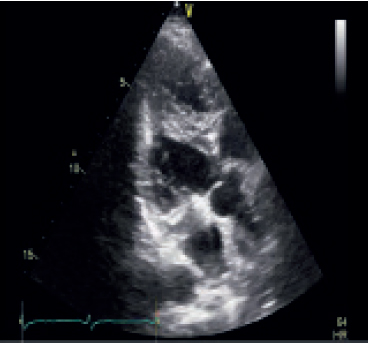
Figure 2. Apical long axis view showing the prominent muscle band extending from apical free wall of the LV to basal anterior septum.
Figure 1. Parasternal long axis view with hypertrophied muscle band.
Figure 2. Apical long axis view showing the prominent muscle band extending from apical free wall of the LV to basal anterior septum.
Discussion
False tendons are thin fibrous or fibromuscular bands that traverse the cavity of left ventricle with no connection to the valvular cusps. These can be commonly seen in LV cavity but HMBs are rare. They usually have a benign course, show no progression even in case of large HMBs (1,2) and ECG changes are limited to repolarization abnormalities, which are produced by repolarization of the muscle trunk. A large HMB, which is close to LV apex, can be misinterpreted as apical hypertrophic cardiomyopathy (AHC). However patients with AHC may experience angina or palpitations and it is usually associated with typical ECG changes. LV ventriculography and contrast echocardiography can be helpful in distinguishing between these two. Another differential diagnosis is LV mass (Fig. 3) in which cardiac MRI can help in tissue characterization. In our case, the patient was totally asymptomatic and there were no ECG changes and no evidence of arrhythmic activity on Holter. Our patient was advised periodic followup with ECG and echocardiography monitoring.
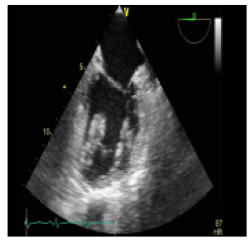
Figure 3. Apical two chamber view showing the hypertrophied muscle band appearing like a mass arising from the apex.
Figure 3. Apical two chamber view showing the hypertrophied muscle band appearing like a mass arising from the apex.
Conclusions
HMB in LV is a rare but benign entity. Patient can be totally asymptomatic or can have atypical chest pain with intermittent palpitations with ECG changes varying from T-wave inversion in limb and precordial leads with junctional rhythm to absolutely normal ECG. HMB can be confused with AHC and LV mass. Detailed echocardiography with or without contrast and MRI can help in differentiating these entities.
References
- Salazar J. Left ventricular anomalous muscle band and electrocardiographic repolarization changes. Pediatr Cardiol. 1997;18:434–6.
- Sutton MG, Dubrey S, Oldershaw PJ. Muscular false tendons, aberrant left ventricular papillary musculature, and severe electrocardiographic repolarisation abnormalities: A new syndrome. Br Heart J. 1994;71:187–90.
- Joo CU, Kim MS, Park JH. Left ventricular anomalous muscle band and electrocardiographic repolarization changes in a 5 year old girl. Korean Circulation J. 2005;35:934–6.
- Lee SH, Ryu HM, Lee JH, Lee H, Park SH, Bae MH, Yang DH. A case of an anomalous hypertrophied muscle band in the left ventricle. J Cardiovasc Ultrasound. 2012;20:97–9.
- Bhairappa S, Mahla H, Kumar SK, Manjunath CN. Anomalous hypertrophied muscle band in LV mimicking LV mass. BMJ Case Rep. 2013 Dec 17;2013. pii: bcr-2013-200819.
