The Journal of Clinical and Preventive Cardiology has moved to a new website. You are currently visiting the old
website of the journal. To access the latest content, please visit www.jcpconline.org.
Effect of Tricuspid Stenosis on Tricuspid Anulus Tissue Doppler Imaging Velocities
Volume 2, Jan 2013
Sita Ram Mittal, DM, Ajmer, India
J Clin Prev Cardiol. 2013;2(1):53-4
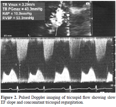
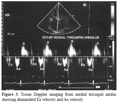
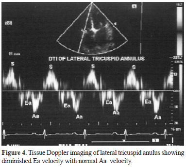
A 20-year-old female was referred for echocardiography. Echocardiography showed tight mitral stenosis (MS) with pliable leaflets. Tricuspid leaflets were thick and showed diastolic doming, diagnostic of tricuspid stenosis (TS) (Figure 1). There was no calcification. Pulsed Doppler evaluation of tricuspid flow revealed slow E-F slope with moderate tricuspid regurgitation (Figure 2). Tissue Doppler imaging revealed reduction in early diastolic velocity (Ea) of lateral as well as medial tricuspid anulus (Figures 3, 4). Late diastolic velocity (Aa) velocity was not increased.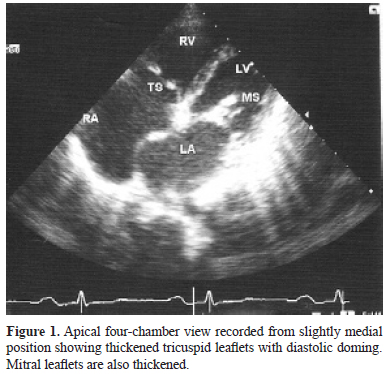
Decreased and slow filling of right ventricle due to TS probably contributed to decreased velocity of Ea wave of tricuspid anulus. Right atrium cannot fill the right ventricle rapidly due to TS. This could contribute to normal or reduced Aa velocity of tricuspid anulus.
Pulmonary artery hypertension secondary to MS could affect right ventricular diastolic functions and reduce Ea
velocity. Pulmonary artery hypertension is, however, associated with increased Aa velocity which was not the
case in our patient. Calcification of anulus can also cause decreased movement. Rheumatic myocarditis could
affect right ventricle and could contribute to decreased Ea velocity. Such a pathology is, however, likely, to
increase Aa velocity. Subvalvular pathology and ball valve thrombus can also affect tissue Doppler velocities in this scenario. Significant tricuspid regurgitation may also change angle of incidence between systole and diastole in such a scenario.
Grant Support
None
Source of Funding
None
Conflict of Interest
None
