The Journal of Clinical and Preventive Cardiology has moved to a new website. You are currently visiting the old
website of the journal. To access the latest content, please visit www.jcpconline.org.
Comparative Diagnostic Accuracy of Different Measures of Preclinical Atherosclerosis for Detection of Atherosclerotic Coronary Artery Disease
Volume 3, Apr 2014
Ravi R Kasliwal MD, DM, Manish Bansal MD, DNB, Rahul Mehrotra MD, DNB, Kulbir Ahlawat, MD, Naresh Trehan MD, Gurgaon, India
J Clin Prev Caridiol. 2014;3(2):36-42
Introduction
Early detection of atherosclerosis, when still in its preclinical stage, has emerged as a promising approach to facilitate optimum cardiovascular (CV) risk stratification of asymptomatic individuals (1–4). Although definitive outcome data is lacking, preliminary evidence suggests that addition of atherosclerosis imaging to conventional risk assessment tools may help better determine the nature and aggressiveness of the preventive measures and can thus improve clinical outcomes (5). In addition, limited evidence also suggests that detection of structural evidence of atherosclerosis may improvepatient compliance toward therapeutic measures (6–10).
Numerous tools have been developed for preclinical atherosclerosis assessment, such as carotid intima media thickness (CIMT), brachial artery flow-mediated dilatation, pulse wave velocity (PWV), coronary calcium score (CCS), etc. Although all these techniques detect one or other manifestation of atherosclerotic process, they have substantial methodological differences that are responsible for the differences in their availability, cost, ease of use, repeatability, radiation exposure, etc. Given these differences, it is imperative to determine their relative diagnostic accuracy in order to be able to define their role in routine clinical practice. Although studies conducted in western populations have suggested CCS to have the best predictive accuracy (11,12), only very few studies have compared different modalities of preclinical atherosclerosis detection in Indians (13). The present study was therefore conducted to compare the three commonly used and approved measures of preclinical atherosclerosis assessment – CIMT, PWV and CCS – for the strength of their association with coronary atherosclerosis.
Numerous tools have been developed for preclinical atherosclerosis assessment, such as carotid intima media thickness (CIMT), brachial artery flow-mediated dilatation, pulse wave velocity (PWV), coronary calcium score (CCS), etc. Although all these techniques detect one or other manifestation of atherosclerotic process, they have substantial methodological differences that are responsible for the differences in their availability, cost, ease of use, repeatability, radiation exposure, etc. Given these differences, it is imperative to determine their relative diagnostic accuracy in order to be able to define their role in routine clinical practice. Although studies conducted in western populations have suggested CCS to have the best predictive accuracy (11,12), only very few studies have compared different modalities of preclinical atherosclerosis detection in Indians (13). The present study was therefore conducted to compare the three commonly used and approved measures of preclinical atherosclerosis assessment – CIMT, PWV and CCS – for the strength of their association with coronary atherosclerosis.
Methods
The study included 219 subjects with or without preexisting CV disease (CVD) who had attended CVD prevention clinic at a tertiary care center in North India. The patients were divided into two groups: Non-CAD group, including those without any evidence of atherosclerotic coronary vascular disease (n=124) and CAD group, which included patients with evidence of atherosclerotic coronary vascular disease (n=95). The patients were included in Non-CAD group if they met all the following criteria:
For the purpose of the present study, HT was defined according to Joint National Committee (JNC) 7 guidelines as systolic BP ≥140 mmHg or diastolic BP ≥90 mmHg or previous history of HT or self-reported use of anti-hypertensive medications (14). Diabetes mellitus was defined as fasting blood glucose >126 mg/dL or 2-hour postprandial blood glucose >200 mg/dL or pharmacological treatment for diabetes or previous history of diabetes mellitus. Family history was considered positive if a coronary event had occurred in a male first-degree relative before the age of 55 years or a female first-degree relative before the age of 65 years. Smoking or tobacco use in any form during the preceding month was also considered to be a CV risk factor.
- No previous history of documented CAD and
- No atherosclerotic plaque seen on conventional or computed tomographic (CT) coronary angiography.
In contrast, the patients were assigned to CAD group if any of the following conditions was present: - Any previous history of documented CAD, with or without coronary revascularization,
- Coronary angiography (conventional or CT) showing evidence of significant CAD defined as >50% luminal narrowing involving at least one epicardial coronary artery, or
- Evidence of coronary plaques, even if causing less significant luminal obstruction (<50%), on conventional or CT coronary angiography.
For the purpose of the present study, HT was defined according to Joint National Committee (JNC) 7 guidelines as systolic BP ≥140 mmHg or diastolic BP ≥90 mmHg or previous history of HT or self-reported use of anti-hypertensive medications (14). Diabetes mellitus was defined as fasting blood glucose >126 mg/dL or 2-hour postprandial blood glucose >200 mg/dL or pharmacological treatment for diabetes or previous history of diabetes mellitus. Family history was considered positive if a coronary event had occurred in a male first-degree relative before the age of 55 years or a female first-degree relative before the age of 65 years. Smoking or tobacco use in any form during the preceding month was also considered to be a CV risk factor.
CIMT assessment (Fig. 1) (4)
For CIMT measurement, distal common carotid artery (CCA) was imaged on both sides with a 7.5 MHz frequency linear array transducer, attached to any standard vascular ultrasound machine. The artery was imaged in a longitudinal plane to obtain optimal angle of incidence, defined as the plane in which the bifurcation of the carotid bulb into the internal and external carotid arteries can be visualized simultaneously with the bulb and distal CCA (also known as “tuning fork” view, Fig. 1). Once this view was obtained, finer adjustments in the transducer position were to be done to ensure distal CCA was perfectly horizontal on the screen and “double lines” of intima and adventitia were clearly visualized in the far wall of the CCA (“double-line” sign, Fig. 1). From this view, the CIMT was measured as the distance between lumen-
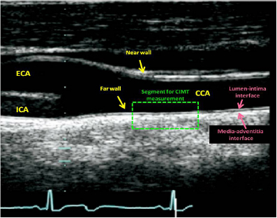
Figure 1. Ultrasound image of distal common carotid artery showing carotid bifurcation in a typical “tuning fork” configuration. The “double line” sign produced because of highly echogenic intima and adventitia with echolucent media lying in between the two is also seen. CCA, common carotid artery; CIMT, carotid intima-media thickness; ECA, external carotid artery; ICA, internal carotid artery.
Figure 2. PeriScope® result sheet showing oscillometric pressure waveforms from different arteries and the various arterial stiffness parameters derived from these pressure waveforms.morning dose was delayed until completion of the test. The procedure was performed in supine position. After 10 minutes of supine rest, four BP cuffs, which were connected to the PeriScope® device, were tied around both arms and both ankles. These BP cuffs carried oscillometric sensors to record pressure waveforms from the underlying arteries. ECG electrodes were applied on wrists and ankles to record ECG simultaneously. The machine then automatically inflated and deflated all the cuffs simultaneously, while recording pressure waveforms from all the four sites. From these pressure waveforms, right and left brachial-ankle PWV (baPWV) were calculated and used for analysis.
Figure 1. Ultrasound image of distal common carotid artery showing carotid bifurcation in a typical “tuning fork” configuration. The “double line” sign produced because of highly echogenic intima and adventitia with echolucent media lying in between the two is also seen. CCA, common carotid artery; CIMT, carotid intima-media thickness; ECA, external carotid artery; ICA, internal carotid artery.
Intima interface and the media-adventitia interface. Plaques, defined as >50% localized thickening of the intima compared to the rest of the wall, were included in the measurement of CIMT if present within the distal 1 cm of CCA.
The CCA was then imaged from two additional complimentary angles, approximately 45° anterior and posterior to the first image and the CIMT measurements were performed. The three values thus obtained for each side were averaged and used for analysis.
Reproducibility of CIMT measurement in our lab has already been documented previously (15).
Measurement of PWV (Fig. 2)
Measurement of PWV was performed using the PeriScope® device (Genesis Medical Systems Pvt Ltd, Hyderabad, India) which has been shown to have high degree of reproducibility for this purpose (16). This device is based on oscillometric method and records arterial pressure waveforms noninvasively. ECG-gated pressure waveforms are recorded simultaneously from both arms and both ankles. From these pressure waveforms, in-built software automatically calculates PWV for different vascular segments (17).
The procedure was performed in the morning, after 10 hours of overnight fast. Participants were asked to refrain from smoking for at least 4 hours before the procedure. Ongoing medications were not discontinued but the
.jpg)
Figure 2. PeriScope® result sheet showing oscillometric pressure waveforms from different arteries and the various arterial stiffness parameters derived from these pressure waveforms.morning dose was delayed until completion of the test. The procedure was performed in supine position. After 10 minutes of supine rest, four BP cuffs, which were connected to the PeriScope® device, were tied around both arms and both ankles. These BP cuffs carried oscillometric sensors to record pressure waveforms from the underlying arteries. ECG electrodes were applied on wrists and ankles to record ECG simultaneously. The machine then automatically inflated and deflated all the cuffs simultaneously, while recording pressure waveforms from all the four sites. From these pressure waveforms, right and left brachial-ankle PWV (baPWV) were calculated and used for analysis.
Coronary calcium scoring
Multidetector CT of the heart was performed using dual-source dual-energy Somatom Definition Flash (Siemens Healthcare, Germany) with 128×0.6mm collimation, rotation time 75 ms and tube voltage of 80 & 140 kV. In single breath-hold, images were acquired from the level of tracheal bifurcation to the base of the heart using prospective ECG triggering with the center of the acquisition at 70% of the R-R interval. From the raw data, the images were reconstructed with standard kernel in 3 mm thick axial, nonoverlapping slices and 25 cm field of view. All image analyses were performed on a dedicated workstation (MMWP and Syngovia Siemens, Germany). A coronary calcified lesion was defined as an area with a density >130 Hounsfield units and covering at least 6 pixels. The Agatston method was used to determine the CCS by multiplying each lesion area by a weighted CT attenuation score in the lesion (18).
Statistical Analysis
The data was managed on Microsoft excel spreadsheet (version 2007, Microsoft Corp, Seattle, WA, USA). Values were expressed as mean (± standard deviation) or as percentages. Comparisons between the groups were carried out using Student’s unpaired t test or chi-square test as appropriate. Receiver-operating characteristics (ROC) curves were constructed to assess diagnostic accuracy of different measures of preclinical atherosclerosis for the presence of CAD. A p value <0.05 was considered statistically significant. All statistical analyses were done using SPSS for Windows (release 15.0, SPSS Inc, Chicago, IL, USA).
Results
Clinical and biochemical characteristics of the two groups are described in Table 1. As compared to the non-CAD patients, those with CAD were older (45.4±10.2 vs. 54.7±9.2 years, P <0.001) and had higher prevalence of hypertension (60.5% vs. 74.7%, P = 0.03). However, there was no difference between the two groups in the prevalence of diabetes, smoking and family history of premature CAD. The two groups also did not show any difference in body-mass index, systolic or diastolic blood pressure and hsCRP. However, as expected, the CAD patients were more likely to be on statin therapy (50.0% vs. 27.6%, P <0.001) which probably accounted for lower total cholesterol, low-density lipoprotein cholesterol and triglyceride levels in them.
Table 1. Clinical and biochemical characteristics of the study population
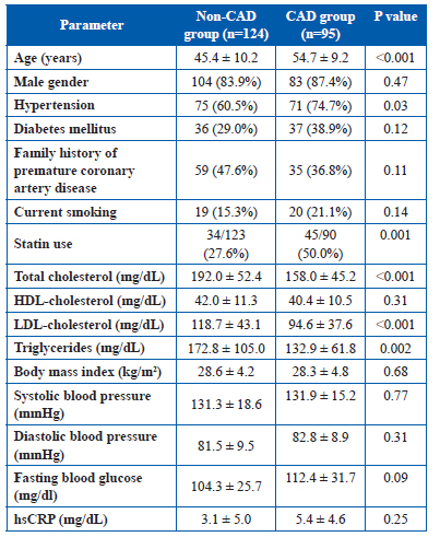
Table 1. Clinical and biochemical characteristics of the study population
All values are in mean ± SD for continuous variables and actual value with percentage in parentheses for categorical variables.
HDL, high-density lipoprotein; hsCRP, high-sensitivity C-reactive protein; LDL, low-density lipoprotein.
Measures of preclinical atherosclerosis (Table 2)
The CAD patients had increased CIMT values on both sides (right side 0.74 ± 0.13 vs. 0.66 ± 0.19 mm; left side 0.79 ± 0.26 vs. 0.67 ± 0.19 mm, and average 0.77 ± 0.18 vs. 0.67 ± 0.18 mm; all P values <0.01). These patients also had significantly increased baPWV on left side (1603.9 ± 381.1 vs. 1414.8 ± 399.0 cm/sec, P <0.0009) but there was no significant difference in right side baPWV or average of the two.
Table 2. Measures of preclinical atherosclerosis in the study population
All values are in mean ± SD for continuous variables.
Table 2. Measures of preclinical atherosclerosis in the study population
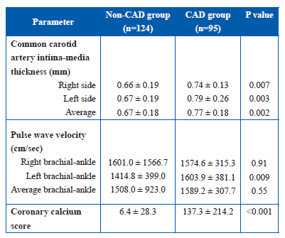
All values are in mean ± SD for continuous variables.
The overall CCS was significantly increased in CAD patients as compared to the patients without CAD (137.3
± 214.2 vs. 6.4 ± 28.3, P <0.001). Among the non-CAD patients, 80.6% patients had 0 score as compared to only 13.3% in the CAD group (P <0.001). In contrast, 31.7% of the CAD patients had CCS >100 whereas only 0.9% of those without CAD had this high CCS (P <0.001).
Similar results were obtained when patients with only significant coronary plaques (causing 50% or greater luminal narrowing) were included in the CAD group.
Diagnostic accuracy of different measures of preclinical atherosclerosis (Fig. 3) Figure 3 displays ROC curves depicting diagnostic accuracy of average CIMT, left baPWV and CCS for the presence of CAD. As shown in the figure, CCS had much greater accuracy (area-under-the-curve 0.89) as compared to CIMT or baPWV (areas-under-the-curve
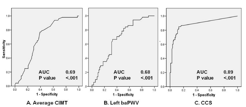 Figure 3. Receiver-operating characteristics curves to demonstrate diagnostic accuracy of different measures of preclinical atherosclerosis for the presence of atherosclerotic coronary vascular disease. (A) Average common carotid artery intima-media thickness (CIMT). (B) Left brachial ankle pulse-wave velocity (baPWV). (C) Coronary calcium score (CCS). AUC, area under the curve.
0.69 and 0.68 respectively, P <0.001). The optimum cutoff value for CCS was 4.05 which resulted in a sensitivity of 85% and specificity of 87%. In comparison, for average CIMT, the cutoff value was 0.65 mm with a sensitivity of 77% and specificity of 61% for diagnosing CAD. The optimum cutoff value for left baPWV was 1453 cm/sec which had a sensitivity and specificity of 67% each. Combining CIMT and baPWV resulted in significant improvement in diagnostic accuracy. Presence of abnormality in at least one of these two parameters (average CIMT >0.65 mm and/or left baPWV >1453 cm/sec) had a sensitivity of 91% for diagnosing CAD and a normal value of both average CIMT and left baPWV could exclude coronary atherosclerosis with 91% certainty (i.e., negative predictive value). Conversely, abnormality in both parameters was associated with 67% likelihood of finding atherosclerotic disease in the coronary arteries.
Figure 3. Receiver-operating characteristics curves to demonstrate diagnostic accuracy of different measures of preclinical atherosclerosis for the presence of atherosclerotic coronary vascular disease. (A) Average common carotid artery intima-media thickness (CIMT). (B) Left brachial ankle pulse-wave velocity (baPWV). (C) Coronary calcium score (CCS). AUC, area under the curve.0.69 and 0.68 respectively, P <0.001). The optimum cutoff value for CCS was 4.05 which resulted in a sensitivity of 85% and specificity of 87%. In comparison, for average CIMT, the cutoff value was 0.65 mm with a sensitivity of 77% and specificity of 61% for diagnosing CAD. The optimum cutoff value for left baPWV was 1453 cm/sec which had a sensitivity and specificity of 67% each. Combining CIMT and baPWV resulted in significant improvement in diagnostic accuracy. Presence of abnormality in at least one of these two parameters (average CIMT >0.65 mm and/or left baPWV >1453 cm/sec) had a sensitivity of 91% for diagnosing CAD and a normal value of both average CIMT and left baPWV could exclude coronary atherosclerosis with 91% certainty (i.e., negative predictive value). Conversely, abnormality in both parameters was associated with 67% likelihood of finding atherosclerotic disease in the coronary arteries.
Discussion
Over the past few decades, atherosclerotic CVD has become the leading cause of death worldwide. The rapid increase in the incidence and prevalence of CVD has led to a growing recognition of the need to develop and implement effective strategies for its prevention. While increasing the overall awareness about the menace of CVD, the reasons for its occurrence and the role of healthy life-style in its prevention appears to be the most effective strategy for CVD prevention, it is greatly limited by the widespread negligence and general apathy of the common public toward healthy life-style measures. A potentially more effective strategy is to identify “high-risk” individuals and then focus all efforts and resources to prevent the development of disease in them.
At present, CV risk assessment is generally performed using conventional CV risk factors and risk algorithms such as Framingham risk score. However, numerous studies have shown that although these risk algorithms perform reasonably well at the population level, their accuracy at the individual level is rather dismal (19–22). Consequently, often there are individuals with multiple CV risk factors and high estimated CV risk who do not develop clinical CVD even in the long term, whereas there are many other patients who present with acute CV event despite being free of all known major CV risk factors. To overcome this limitation, several diagnostic tools have been developed that have the ability to detect atherosclerotic vascular disease in its early, pre-clinical stage and therefore can provide direct evidence of ongoing atherosclerotic process, irrespective of the presence or absence of known CV risk factors. Numerous large-scale studies have unequivocally demonstrated the incremental value of such techniques over conventional CV risk factors in prediction of future CV risk (4,17,23). Although it is yet to be proven that incorporation of these diagnostic techniques into the standard risk assessment approach can improve clinical outcomes, the evidence so far suggests that such a strategy can be helpful in guiding nature, timing and aggressiveness of anti-atherosclerotic therapy, in improving patient compliance toward these measures and may also help in monitoring response to therapy (1,2,4–10).
CIMT, brachial artery flow-mediated dilatation, PWV assessment, ankle-brachial index and CCS are the most commonly used measures of preclinical atherosclerosis. All these tools aim to detect evidence of atherosclerosis in different vascular beds with the underlying rationale that atherosclerosis is a generalized disease and its presence in one vascular bed indirectly implies involvement, though not necessarily concurrently, of other vascular beds too. However, these techniques are methodologically vastly different from each other and therefore have significant differences in their availability, cost, ease of use, repeatability, radiation exposure, etc. As many of these techniques are already available for clinical use, it is important to determine their relative diagnostic accuracy to allow for their judicious, cost-effective and the most meaningful use in routine clinical practice.
Previous studies have shown CCS to have much greater predictive accuracy for future CV events and to have the greatest incremental value over conventional risk assessment algorithms (11,12). However, no study has compared CCS with the other modalities of preclinical atherosclerosis assessment in Indians; thus our study is the first one to provide such data. Similar to the western populations, we too found CCS to have the greatest predictive accuracy for the presence of CAD.
Although CCS is more accurate in predicting CAD as compared to CIMT or PWV, it is expensive, not readily available and is associated with radiation exposure, which limit its use in routine clinical practice. To this extent, our study has provided an important insight. We found that a combination of CIMT and baPWV could provide excellent diagnostic accuracy and could be used as a simpler, less-expensive and radiation-free approach to initial CV risk stratification of the individuals. If both CIMT and left baPWV are normal, CAD can be excluded with high degree of certainty and no further testing may be needed. In contrast, if both are increased, there is a high likelihood of the presence of coronary atherosclerosis and the patient can be subjected to CCS or stress testing based on their clinical presentation. In the remaining individuals with only one of the parameters being abnormal, further evaluation will have to be individualized according to the presence or absenceof other CV risk factors. Thus a hierarchical approach could be utilized to minimize cost, reduce radiation risk and to make it more suitable for wider clinical use, but at the same time retaining its diagnostic accuracy.
Limitations
Our study had some important limitations that warrant attention. The sample size was relatively small, which precluded detailed assessment of the impact of various CV risk factors on the predictive accuracy of different diagnostic markers. Also, CIMT, baPWV and CCS are predominantly used for prediction of future risk of CV events and not for diagnosing incident CAD. As our study was only a cross-sectional one, it was not possible for us to determine the accuracy of these different tools for prediction of future CV risk. Nonetheless, presence of the association with existing coronary atherosclerosis strongly supports their validity as markers of atherosclerotic vascular disease and their ability to predict future CV events. Finally, a significant proportion of the patients in both the groups were on medications, especially statins which must have altered the relationship between preclinical atherosclerosis and CAD. However, use of statins in CAD patients is likely to have only weakened this relationship and thus only undermined our findings.
Conclusions
The present study shows that among different measures of preclinical atherosclerosis, CCS has the best diagnostic accuracy for the presence of coronary atherosclerosis. However, the combination of CIMT and baPWV has an excellent negative predictive value for atherosclerotic coronary vascular disease and can therefore be used as a simple, noninvasive, less-expensive and radiation-free approach to initial CV risk stratification of the individuals. Those with abnormal CIMT or baPWV can then be subjected to CCS for further refinement of their CV risk.
Acknowledgment
We sincerely thank Mr. Arun Rawat and Mr. Ravinder Negi, clinical research coordinators, for the study, for their immense help in data collection and compilation.
Funding
No external source of funding to be mentioned.
Conflict of Interest
None
References
- Greenland P, Alpert JS, Beller GA, et al. 2010 ACCF/AHA guideline for assessment of cardiovascular risk in asymptomatic adults: A report of the american college of cardiology foundation/american heart association task force on practice guidelines. J Am Coll Cardiol. 2010;56:e50–103.
- Mancia G, De Backer G, Dominiczak A, et al. 2007 guidelines for the management of arterial hypertension: The task force for the management of arterial hypertension of the european society of hypertension (esh) and of the european society of cardiology (ESC). Eur Heart J. 2007;28:1462–536.
- Can atherosclerosis imaging techniques improve the detection of patients at risk for ischemic heart disease? Proceedings of the 34th bethesda conference. Bethesda, Maryland, USA. October 7, 2002. J Am Coll Cardiol. 2003;41:1856–917.
- Stein JH, Korcarz CE, Hurst RT, et al. Use of carotid ultrasound to identify subclinical vascular disease and evaluate cardiovascular disease risk: A consensus statement from the american society of echocardiography carotid intima-media thickness task force endorsed by the society for vascular medicine. J Am Soc Echocardiogr. 2008;21:93–111.
- Shah PK. Screening asymptomatic subjects for subclinical atherosclerosis: Can we, does it matter, and should we? J Am Coll Cardiol. 2010;56:98–105.
- Bovet P, Perret F, Cornuz J, Quilindo J, Paccaud F. Improved smoking cessation in smokers given ultrasound photographs of their own atherosclerotic plaques. Prev Med. 2002;34:215–20.
- Wyman RA, Gimelli G, McBride PE, Korcarz CE, Stein JH. Does detection of carotid plaque affect physician behavior or motivate patients? Am Heart J. 2007;154:1072–7.
- Barth JD. Which tools are in your cardiac workshop? Carotid ultrasound, endothelial function, and magnetic resonance imaging. Am J Cardiol. 2001;87:8A–14A.
- Kalia NK, Miller LG, Nasir K, Blumenthal RS, Agrawal N, Budoff MJ. Visualizing coronary calcium is associated with improvements in adherence to statin therapy. Atherosclerosis. 2006;185:394–9.
- Taylor AJ, Bindeman J, Feuerstein I, et al. Community-based provision of statin and aspirin after the detection of coronary artery calcium within a community-based screening cohort. J Am Coll Cardiol. 2008;51:1337–41.
- Folsom AR, Kronmal RA, Detrano RC, et al. Coronary artery calcification compared with carotid intima-media thickness in the prediction of cardiovascular disease incidence: The multi-ethnic study of atherosclerosis (mesa). Arch Intern Med. 2008;168:1333–9.
- Simon A, Chironi G, Levenson J. Comparative performance of subclinical atherosclerosis tests in predicting coronary heart disease in asymptomatic individuals. Eur Heart J. 2007;28:2967–71.
- Kasliwal RR, Bansal M, Bhargava K, Gupta H, Tandon S, Agrawal V. Carotid intima-media thickness and brachial-ankle pulse wave velocity in patients with and without coronary artery disease. Indian Heart J. 2004;56:117–22.
- Chobanian AV, Bakris GL, Black HR, et al. The seventh report of the joint national committee on prevention, detection, evaluation, and treatment of high blood pressure: The JNC 7 report. JAMA. 2003;289:2560–72.
- Kasliwal RR, Agrawal S, Bansal M. Carotid intima-media thickness and brachial artery flow-mediated dilatation in patients with and without metabolic syndrome. Indian Heart J. 2006;58:42–6.
- Naidu MU, Reddy BM, Yashmaina S, Patnaik AN, Rani PU. Validity and reproducibility of arterial pulse wave velocity measurement using new device with oscillometric technique: A pilot study. Biomed Eng Online. 2005;4:49.
- Laurent S, Cockcroft J, Van Bortel L, et al. Expert consensus document on arterial stiffness: Methodological issues and clinical applications. Eur Heart J. 2006;27:2588–605.
- Agatston AS, Janowitz WR, Hildner FJ, Zusmer NR, Viamonte M, Jr., Detrano R. Quantification of coronary artery calcium using ultrafast computed tomography. J Am Coll Cardiol. 1990;15:827–32.
- Nasir K, Michos ED, Blumenthal RS, Raggi P. Detection of high-risk young adults and women by coronary calcium and national cholesterol education program panel III guidelines. J Am Coll Cardiol. 2005;46:1931–6.
- Lauer MS. Primary prevention of atherosclerotic cardiovascular disease: The high public burden of low individual risk. JAMA. 2007;297:1376–8.
- Shaw LJ, Blumenthal RS, Raggi P. Screening asymptomatic low-risk individuals for coronary heart disease: Issues and controversies. J Nucl Cardiol. 2004;11:382–7.
- Akosah KO, Schaper A, Cogbill C, Schoenfeld P. Preventing myocardial infarction in the young adult in the first place: How do the national cholesterol education panel III guidelines perform? J Am Coll Cardiol. 2003;41:1475–9.
- Greenland P, Bonow RO, Brundage BH, et al. ACCF/AHA 2007 clinical expert consensus document on coronary artery calcium scoring by computed tomography in global cardiovascular risk assessment and in evaluation of patients with chest pain: A report of the american college of cardiology foundation clinical expert consensus task force (ACCF/AHA writing committee to update the 2000 expert consensus document on electron beam computed tomography) developed in collaboration with the society of atherosclerosis imaging and prevention and the society of cardiovascular computed tomography. J Am Coll Cardiol. 2007;49:378–402.
